- Article
- Source: Campus Sanofi
- 23 Oct 2023
What is Gaucher disease?
Gaucher disease is one of the most common lysosomal storage disorders, affecting an estimated 1 in 40,000 to 1 in 100,000 people around the world.1 It can be diagnosed at any age from infancy to late adulthood. It is an inherited deficiency of the lysosomal enzyme acid-β-glucosidase (glucocerebrosidase, GBA1), which results in the accumulation of glucocerebroside within lysosomes of macrophages.1
.jpg)
Gaucher disease can be classified into three types, which make a phenotypic continuum ranging from mild to severe nervous symptoms.2–4 The classic categories of types 1, 2, and 3 have blurred edges along the continuum of the disease.
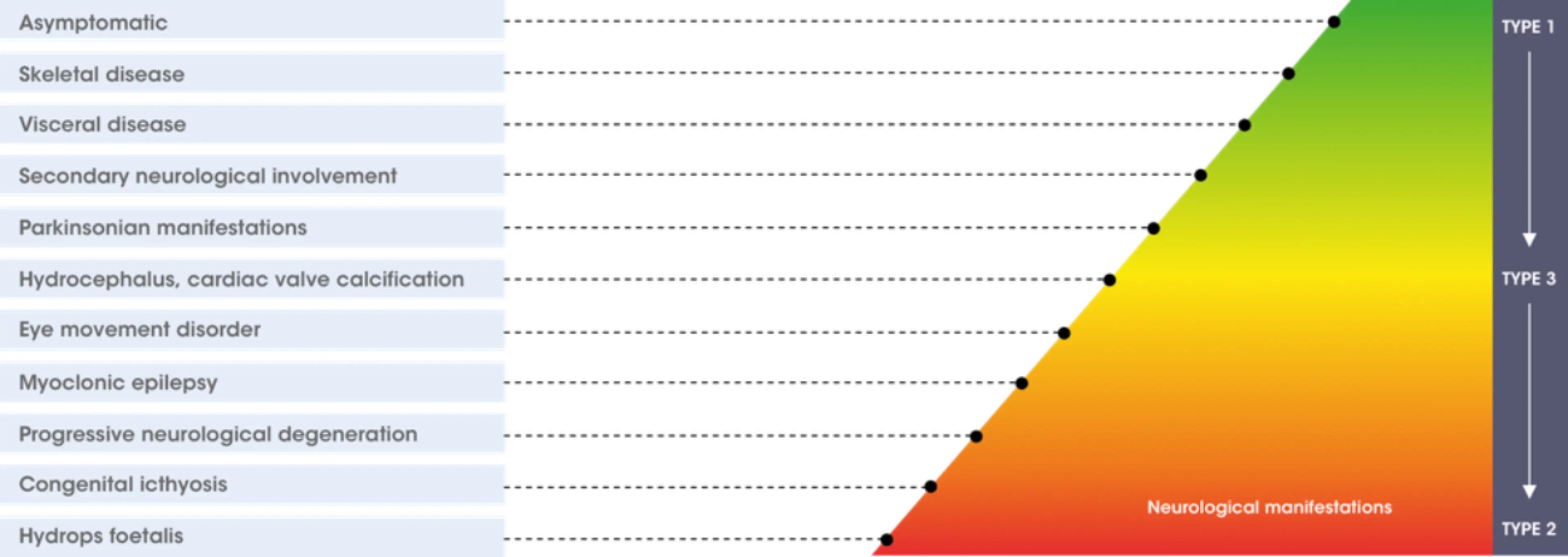
Adapted from Sidransky E, 2004. Mol Genet Metab. 83(1–2):6–15.3
The table summarises aspects of Gaucher disease according to the three different types.1,5
| Type 1 Non neuronopathic(≥1/100 to <1/10) | Type 2 Acute neuronopathic (≥1/100 to <1/10) | Type 3 Chronic neuronopathic (≥1/100 to <1/10) | |
| Prevalence | 1:50,000 – 1:100,000 (pan-ethnic) 1:850 (Ashkenazi Jews) | < 1:150,000 (pan-ethnic) | < 1:150,000 (pan-ethnic) |
| Age at presentation | Any | Infancy | Childhood |
| Lifespan | Variable | < 2 y | < 40 y |
| Primary CNS disease | None | Severe | Mild to severe |
| Hepatosplenomegaly | Mild to severe | Severe | Mild to severe |
| Haematologic abnormalities | Mild to severe | Severe | Mild to severe |
| Osseous symptoms | Mild to severe | None | Mild to moderate |
References
- Mistry PK, et al. Am J Hematol 2011. 86(1):110–115.
- Charrow J, et al. Clin Genet 2007. 71(3):211–215.
- Sidransky E, et al. Mol Genet Metab. 2004. 83(1–2):6–15.
- Sidransky E, et al. Gaucher disease clinical presentation. Updated November
- Niederau C. Gaucher Disease 3rd edition. Bremen: Uni-Med, 2017.
MAT-XU-2201084 (v2.0) Date of preparation: October 2023