Immunological Evaluation Pre and Post Transplant

- In this presentation, Dr. Sukant Kishore Das, a consultant nephrologist with 15 years of experience in Bhubaneswar, specializing in kidney transplantation and interventional nephrology, focuses on pre- and post-transplant immunological evaluation, covering key advancements and challenges.
- Transplant immunology has evolved significantly, improving patient outcomes and graft survival. However, major challenges remain, particularly in graft rejection diagnosis, donor-specific antibody detection, and individualized immunosuppression. Here, we will discuss critical aspects, including HLA typing, ABO compatibility, virtual cross-matching, and epitope analysis, which are crucial for minimizing rejection risks and optimizing transplant success.
- This session is designed for physicians and nephrology fellows to explore the latest updates in pre- and post-transplant immunology, aiming for improved kidney transplant outcomes through better diagnostics and immunosuppressive strategies.
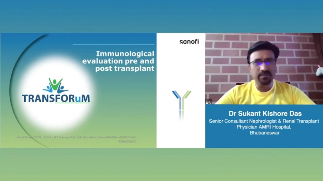
Dr. Sukant Kishore Das
Hello everyone, this is Dr. Sukant Kishore Das. As you all know, I am a consultant in nephrology and I am a DM nephrologist from SZPHI back almost 15 years back and since then I've been practicing in Bhubaneswar in different hospitals as a nephrologist and a transplant physician. And I have had the opportunity to be an organizing secretary of ISN ISKCON 2022 which is the East-Jungle Conference of Indian Society of Nephrology and of course I have been actively busy with doing various publications in the field of kidney nephrology and especially in the area of transplantation and my core area of interest is kidney transplantation as well as interventional nephrology. So today we will be talking about immunological evaluation pre and post transplantation. So I welcome all of you to this session which is going to be the traditional areas of pre-transplant immunological evaluation as well as exciting areas in the field of kidney transplantation and pre-transplantation immunobiology. Let me re-emphasize once again this is meant for physicians and as well as basically fellows of nephrology and kidney transplantation who are interested in various immunological pre and post transplantation and what are the latest updates related to this. Now transplant immunology has made significant improvements and progresses over the last 20 years. There have been certain areas in transplant immunology which of course need to be looked at greater perspective. One is of course it improves overall kidney transplantation outcome, the patient's quality of life, the survival of end-stage renal disease in patients as well as the diagnosis and treatment of graft rejection. Diagnosis and treatment of graft rejection is the most important area where it needs to be highlighted and that is the end point towards which all our efforts should be oriented because we need to minimize the instances of graft rejection and graft failure. Now this targets not just T cell rejection, not just B cell rejection, but it targets the entire area of graft rejection. We have made significant progresses. We have made significant progresses in terms of anti-HLA antibodies and donor-specific antibodies as well, but we still need substantial progress. Where we need this substantial progress? We need in terms of non-invasive biomarkers. We still do not have a very clinically proven reliable non-invasive biomarker which can work as one point marker to predict and not just predict pre-transplant, but also in a post-transplantation patient, how far the rejection is there and can be a definitive marker of active rejection. Till now, we only have kidney biopsy as a definitive marker of active rejection. Then there is continued reduction or immunosuppression. Can we plan for something in individualized immunosuppression in this patient? How we can plan for individualized immunosuppression and how we can reduce the cross immunosuppression in a patient and overall reduce the chances of infections in this patient, reduce the chances of cross immunosuppression status of infection and improve the overall survival of these patients. Now the next thing we will be focusing on, now let us discuss the basics of immunological testing in kidney transplantation, which include testing for ABO blood group compatibility and the testing for HLA typing and donor-specific antibodies. You know, testing for ABO incompatibility basically focuses on three important things. Two methods used to reduce circulating ABO antibody titers. So, first of all, you have to measure the titers, then reduce the cross antibody titers by two different methods. And also, one has to look at the B cell population reduction by methods such as rituximab or splenectomy. Splenectomy has become gradually obsolete as ever since rituximab has taken its place and rituximab is increasingly used. Even lesser and lesser doses of rituximab are used these days, which have significantly replaced and almost replaced the robot splenectomy. And the goal in ABO incompatibility transplantation is to achieve titers less than 1 is to 8 to 1 is to 13 before proceeding for a viable kidney transplantation. Now according to the next important area where we need to focus is HLA typing. HLA mismatch offers affects long-term graft survival, not just at the resection. In fact, it is even more challenging in areas of ABO incompatible transplantation because HLA mismatches actually are not good for graft survival. A 5-year graft survival for diseased donor kidney transplants is 77% with 0 HLA mismatch and it was 67% with 6 HLA mismatches. DSS, what are DSS? DSS are donor-specific antibodies. They are recipient's antibodies specific to the donor. High titers are contraindication for kidney transplantation and mean fluorescent intensity is a major of donor-specific antibodies in the recipient via the transplantation. And then the strength of mean fluorescence MFI index is mean fluorescence intensity is an is an index of how significant the antibody mediated resection is going to be. So one to look at desensitization of pathogenic DSS using plasmapheresis, rituximab, IVIGP transplantation and remember more than 5000 MFI is a contraindication to kidney transplantation. Now looking at HLA antigens, comparison of HLA antigens at a molecular level. Here we have a figure, this represents two sets of HLA's between the HLA of the recipient and HLA of the donor. Now remember it's not just the cross HLA, one has to look at the HLA structure at a molecular level. Now traditional HLA full antigen mismatches only evaluate whether the donor and the recipient molecules are the same or the different. We just look at whether it's HLA-A one in one molecule, HLA-A 11 and HLA-DR one in one and HLA-DR 11 in another is stated in this example. So traditional HLA whole antigen mismatches only evaluate whether the donor and the recipient molecules are the same or different. The issue is that some mismatches of HLA are nearly identical while others may be very disparate, while others may be very disparate and the information ignored while traditional HLA mismatches are carried out. But this relative difference can be captured and quantified by HLA molecular mismatch comparisons. In this example one and we have the recipient who has got HLA-DR one and the donor has got HLA-DR 11. They are of course clearly different but when we go for a molecular comparison it was found that HLA donors shared three out of five HLA epilates with the recipient. This means that the donor had two mismatched epilates that were different from those in the recipient's HLA antigens. So epilate is the new area which needs to be targeted. Now conventional methods to detect HLA antibodies cell-based assays were used. There were CDC cross match, the screen for preformed antibodies recipients. CDC is one of the still popular methods and of course one of the must-to-be-used methods as a pre-transplant antibody assessment. It can be retransplant or C-cell cross match. In a T-cell cross match T lymphocytes are isolated from the donor. They are mixed with serum of the recipient. Now recognize HLA-1 and bind to antibody in the recipients. When the complement is added the cells undergo lyses if antibodies are present. Now dye penetrates into the lysed cells and can detect dead cells. If there is no antibody complement will not be deactivated and there will be no cell death. No dye will be taken up due to absence of dead cells. This is the traditional CDC antibody mismatch and CDC antibody test. Now CDC is still one of the most commonly used antibody tests we employ. Definitive tests to be carried out. I mean no transplant center will proceed without CDC negative test assays to be carried out. Conventional methods have to be compared one after another. What are the different other conventional methods to detect HLA antibodies? Apart from CDC, the methods can be ELISA method, the in-flow breed dependent cross matches and Luminex single antigen BPC. Now in ELISA method HLA bound serum detects anti-HLA antibodies with colored product using enzyme level immunopharmacology. These methods always employ some sort of fluorescent method of assessment and color level coded of different enzymes to exactly identify the mismatched HLAs. In in-flow breed dependent cross matches, cross match uses fluorescent HLA bits to detect IgM IgG antibody against both HLA1 and HLA2 with complement fixer cell. And a third and most important area is Luminex uses HLA immobilized microspheres on your cytomembrane to detect antibodies with MFI quantification. The contraindicating MMI MFI being over 5000 for immunotranspiration. Now, once again, let me explain this slide. Once again, the HLA leukocyte, the human leukocyte antigen can be immobilized on a solid surface and results can be detected using three different methods. The first method is enzyme level observant assay, as it's the left side of the slide, which involves incubating HLA bound serum patients on a plastic surface to detect antibodies. If the antibodies bind to the antigen, an enzyme labeled immunoglobulin produces color product. If there are no anti HLA antibodies in the patient's serum, the anti human immunoglobulin G cannot attach and it is washed away and there is no color product. The second, as I have stated in the middle of the slide, is the middle box of the slide presents the in-flow breed dependent cross match, which uses fluorescent latex HLA beads to detect IgM and IgG antibodies again, class one and class two with component fixation, where multiple HLA antigens can be located on the beads. And the third method, the most important and the most recent one is of course, the Luminex single antigen bead assay, which uses HLA molecules immobilized in 100 monosterin microspheres with fluorescent latex. The serum is incubated with microspheres coated with HLA molecules so that antibodies are detected within 4 cycles of growth. And near fluorescence intensity of the patient and MFR value data of the hydrogen is considered a predication of immunosuppression. Next we will look at virtual cross matching and epitope analysis. In virtual cross matching, we have virtual cross match, which assesses donor recipient HLA compatibility using recipient anti-HLA antibodies without donor lymphocytes. It is also known as virtual cross matching, which assesses compatibility between kidney transplant recipients and donors without actually using donor cells. It can be single antigen bead assay or high resolution typing, HLA typing. Now virtual cross match usefulness and safety have been reported, but controversy remains regarding whether it can replace physical cross matching. Now epitope analysis is a method to infer targeted epitope from single antigen bead assay reaction back into a cell to assess HLA compatibility. Donor specific HLA antibody, a key player in hyper-acute rejection. Now after assessing all these things, we have to look at the importance of donor specific antibodies. Look at the relevance and the relevant frequency and clinical frequency and the relevance of donor specific antibodies as low as 4% or it can be as high as 50%. But one has to remember that pre-existing or de novo donor specific antibodies have a deleterious effect on graft output across solid organ transplants. Now HLA antibodies in the occurrence of early adverse events are predicted. Early adverse events do not affect the long-term graft survival. If a good graft function is achieved by the end of the third post-transplant phase. So one has to remember that if we are aware of the pre-existing donor specific antibodies, we can modify the occurrence of hyper-acute rejection or try to avoid the occurrence of hyper-acute rejection. We can minimize acute rejection in the early post-transplant period and thereby we can ensure to have a good post-transplant out. Now the clinical frequency as I have just stated of the donor specific antibodies is 4% to 50%. But this is a well-established fact that pre-existing or de novo DSI has an unfavorable effect on graft outputs. So one has to have a clear-cut idea to minimize its impact in the post-transplantation period and thereby one has to have a tool to assess in the pre-transplant period. Now what are the risk factors for HLA sensitization? We all understand the risk factors. Of course the patient is going to get a second kidney transplantation or the patient has got multiple pregnancies which sensitizes HLA leading to the development of HLA class 1 antibodies and if the patient has kidney transplants or has multiple blood transfusions, despite proper antigen cross-matching in blood transfusion, the mortality is the leading cause. Trally although poorly immunogenic persistent HLA allosensitization requires multiple transfusions. Now why it is so important to look at the history of pre-transplant graft transfusions because there is a leading cause of mortality is transfusion related acute brain injury. Although transfusion is poorly immunogenic persistent HLA allosensitization induces induction requires multiple transfusions. Now association of HLA mismatching with graft survival and mortality. Now HLA mismatching is a critical prognostic factor which affects graft and recipient survival. Now if we specifically look at HLA-DR mismatching and HLA-A mismatching. Now remember substantial impact is contributed by HLA-DR mismatching and the same level of contribution is not HLA-A mismatching which is insignificant impact on graft survival. More HLA mismatches are significantly associated with a greater risk of failure and the mortality. So the important tests to detect the anti-HLA and other preformed antibodies in the plasma serum include cell-based assays and solid-based assays. HLA-DR mismatches were significantly associated in this particular study where 12% higher risk of over-implant failure whereas A mismatch was associated with far less almost 50% less with 6% higher risk. Now look at the role of epilates. What are epilates? Epilates are small. Epilates are particular when we look at a detailed molecular level analysis of the HLA we get the epilates. Epilates are small configurations of surface exposed amino acids on the surface of immune deposit antigens. They are functional units of structural epitopes on donor HLA. They are functional units of structural epitopes of donor HLA. Potentially they are recognized by complementarity determining reasons of the paratope of the recipients B cell receptors and or antibodies and generally determine the specificity of antibody through interaction with complementarity determining reason of the heavy chain of the antibody. Now epilates we know the conventionally the role of epitopes and paratopes and they are recognized by the paratopes of the antibodies. Epilates help to locate donor hormone immune crucial structural differences to the recipients HLA. They play a role a significant role in improving transplant outcomes by decreasing donor specific antibody formation and potentially providing an opportunity to limit the immunosuppression of well matched transplants. HLA epilates is an immunologic unit of HLA antigens as I just stated. Now HLA epitopes and resection in transplantation. HLA antigens are protein molecules found on the surface of cells that have vital function in the immune system's capacity to infection between epitopes, own cells and other cells. The aid in identification of foreign substances helps to trigger immune response against them while not attacking the body's own cells. HLA epilates are specific amino acids that are part of protein chain and are dispersed throughout the HLA antigen structure. These epilates are recognized by immune system as foreign. They are not present in the patient's own HLA antigen. They can trigger a new response leading to cellular immunization of the transplanted patients. Now let's discuss the concept of HLA antigen epilate which is being considered as the new currency for measuring HLA compatibility. Let's discuss this HLA epilate once again. The way HLA compatibility is defined needs to be changed if there is a match which is based on conventional HLA match does not quantify the immunological risk whereas there is a better quantification risk based on HLA epilate use of HLA epilates. Now the current concept of HLA compatibility is inadequate. Why? Because the conventional HLA match does not accurately quantify the risk. There is a need for a better representation and this is where the role of HLA epilate comes. It is considered as the new currency in this HLA compatibility. It provides the most precise measurement of the immunological risk involved in organ transplantation compared to the conventional HLA match. What does it do? It improves risk assessment of the donor resident HLA compatibility. It's the best tool for risk stratification at the population level and epilate's clinical relevance is crucial for tailored kidney transplantation. That's our ultimate aim, tailored kidney transplantation and the DQ epilate mismatches need special mention. It increased de novo DSF formation, graft rejection and failure after kidney transplantation. This is responsible. DQA1 and DQB1 alleles could improve transplant outcomes. Personalized IS guided by antibody verified HLA DQ epilate mismatch load analysis may improve the overall outcome. So incorporating molecular matching for DQA1 and DQB alleles may aid in self-reducing the risk of de novo DSF formation and potentially enhance outcomes of kidney transplantation. Now we will discuss the role of cell-free DNA as an immunological marker. This immunological marker test measures the proportion of total cell-free DNA from the donor and the recipient. Now donor derived cell-free DNA is typically low in concentration and usually less than 1% when there is no active catalyst to the animal. Higher amounts of daily cell-free DNA are released during graft rejection. Early rise of donor derived cell-free DNA during acute rejection have been observed in kidney transplant recipients. So as the donor derived cell-free DNA rises, they are increasingly associated with episodes of acute graft rejection. So if there will be a method of quantitative assessment or quantitative measurement of donor derived cell-free DNAs and their interpretation, this can be used to evaluate acute graft rejection situations. So it will provide as a non-invasive diagnostic tool for antibody mediated rejection. Now we can use donor derived cell-free DNA as well as DSA testing, which will give us an overall improved non-invasive diagnosis of active antibody mediated rejection and patients with donor derived cell-free DNA results have high probability of antibody mediated rejection. We need a multi-parameter pre-transplant prediction of acute cellular rejection. So using new tools, continuous and rank normalized HLA-1 single antigen with reactivity profiles provide high accuracy for risk assessment for pre-transplant acute cellular rejection. Based on that, we can modify which patients are high risk patients and which patients are low risk patients, and this model will give us detection and treatment of rejection and ultimately improves graft survival. So we have got, now there is a new area we will look into. One is trans-skeptomic biomarker. Much of this is an experimental stage. So all these biomarkers, in course of time, may use and may have no longer be confined to textbooks, will have their use increasingly in day-to-day clinical practice. And if you look into further, this trans-skeptomic biomarker has a higher predictive capacity than clinical variables. Epigenetic biomarkers will have more predictive outcomes and detect chronic allograft dysfunction. And proteomic biomarkers are related to AWF-4, predicts poorer graft outcomes. In one way or the other, they can go for identifying the short-term, long-term, and overall survival of the graft. So if we have a, in a nutshell, we have to explain, so how do we have a pre-transplant algorithm for histocompatibility testing? We have to go for the cross-match, whether CDC or pro-cytometry cross-match. If it is negative, go ahead for transplant. If it is positive, then ideally look for donor-specific antibodies. If there are no donor-specific antibodies, and if the pro-cytometry is negative, we can go ahead with transplant. If DSA is present and pro-cytometry is positive, we'll have to go for alternative donor or If DSA is present but pro-cytometry is negative, desensitization or alternative donor, we can adopt desensitization. But remember, even with desensitization, the risk of long-term dysfunction is always there. Now, practical histocompatibility approach in patients with anti-HLA. Anti-HLA screen positive. Look for DSA expensive and, I mean, look for DSA expensive and indecisive. So, do low cross-match less expensive and indecisive. So, if we look for virtual cross-matches, those can be more expensive, but low cross-match is less expensive. Pro-cytometry cross-match negative, transplant is visible. Pro-cytometry cross-match positive, and it depends on the donor-specific antibody. So, clinical practical guidelines, clinical practice guidelines, immunological assessment state, the guidelines. It communicates all immunological assessment in clinical practice. Communicate all sensitizing agents. HLA typing using molecular methods. HLA antibody testing and transplant evaluation. HLA antibody testing and solid phase assessment. HLA typing using molecular methods, not routinely used on HLA antibodies. Not routinely testing for candidates of HLA binding in HLA antibodies. Access to transplantation based on blood type and histoplasticity test results, and antibody avoidance before desensitization. So, with that, I conclude this presentation. You might have had an overall understanding. I know there are several concepts which we do not use regularly as a transplant pathologist because there are too many types of immunology. So, there are certain areas that we have discussed which are already urgent, and there are certain areas which we are discussing which are partly in our use, and there are some areas which are still far-fetched and will be used in subsequent cases. So, with that, I conclude my presentation. Thank you very much.