Assessing Pediatric Lung Function

.2025-10-15-10-02-51.png)
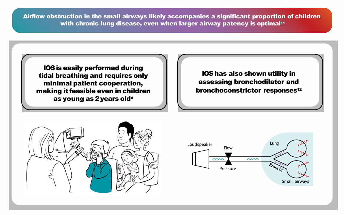
Learn More About These Available Noninvasive Techniques for Assessment of Lung Function3-5

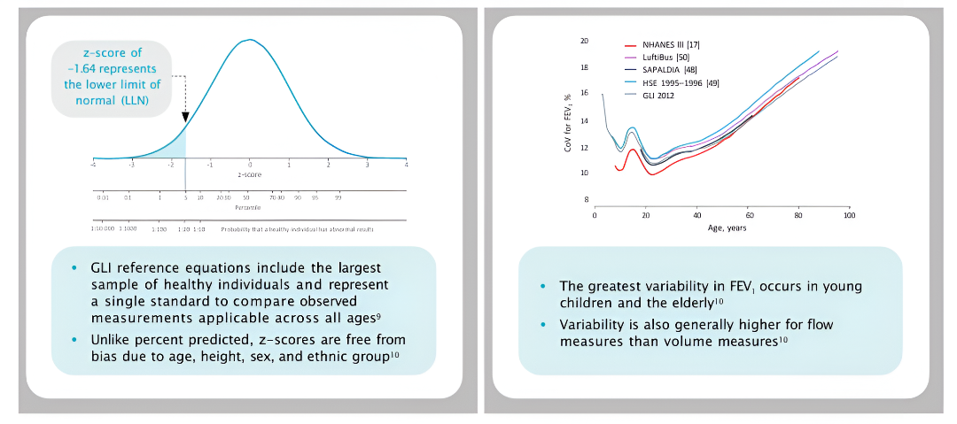
Global Lung Function Initiative (GLI) Suggests Using Z-Scores to Standardize FEV1 Measurements and Minimize Confounding Variables9,10
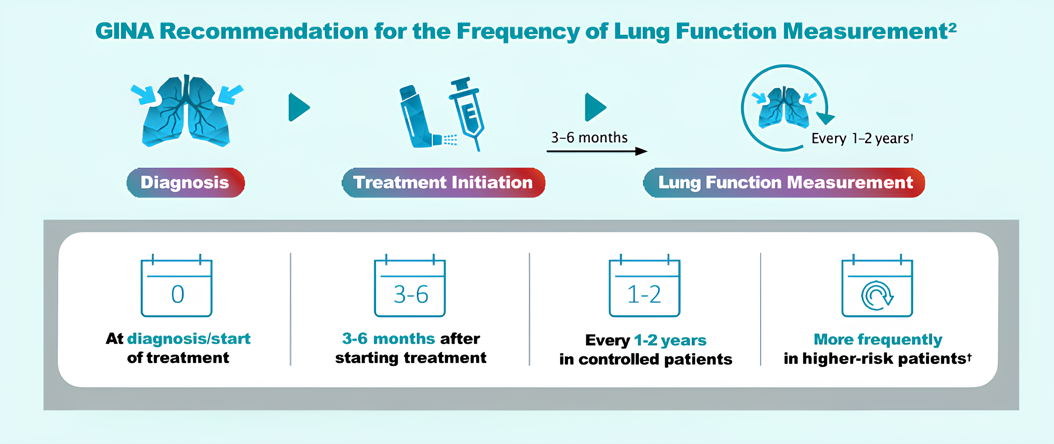
Peak expiratory flow monitoring with a respiratory diary may also sometimes be useful for continued assessment of lung function and earlier detection of future exacerbations outside of a physician’s office2
References:
CoV, coefficient of variation; FEV1, forced expiratory volume in 1 second; FVC, forced vital capacity; GLI, Global Lung Function Initiative; HSE, Health Survey for England; NHANES, National Health and Nutritional Examination Survey; SAPALDIA, Swiss Cohort Study on Air Pollution and Lung and Heart Diseases in Adults.

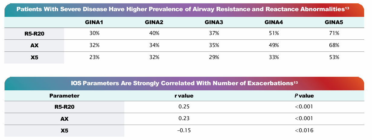
Abnormalities in IOS components (compared to reference values) are more prevalent in individuals with more severe disease according to GINA guidelines and can be used to identify inadequate asthma control in pediatric patients with normal spirometry13,14
References:
AX, area of reactance; GINA, Global Initiative for Asthma; IOS, impulse oscillometry; R5-R20, peripheral airway resistance; X5, reactance at 5Hz.

References:
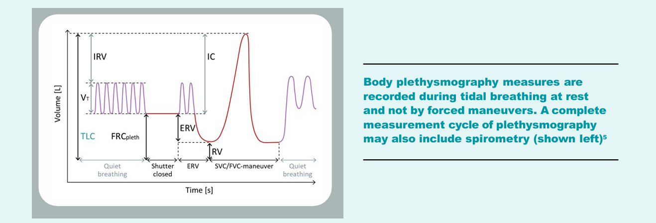
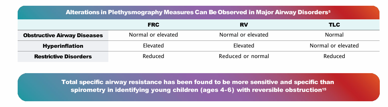
ERV, expiratory reserve volume; FRC, functional residual capacity; FVC, forced vital capacity; IC, inspiratory capacity; IRV, inspiratory reserve volume; RV, residual volume; SVC, slow vital capacity; TLC, total lung capacity; Vt, volume-time.

1. Fuhlbrigge AL, et al. Pediatrics. 2006;118(2):e347-e355.
2. Global Initiative for Asthma (GINA). Global strategy for asthma management and prevention. Updated May 2025. Accessed May 30, 2025. https://ginasthma.org/2025-gina-strategy-report/
3. Merck Manual Professional Version. Airflow, lung volumes, and flow-volume loop. Accessed January 23, 2024. https:// www.merckmanuals.com/professional/pulmonary-disorders/tests-of-pulmonary-function-pft/airflow, lung-volumes,-and-flow-volume-loop
4. Galant SP, et al. Ann Allergy Asthma Immunol. 2017;118(6):664-671.
5. Criée CP, et al. Respir Med. 2011;105(7):959-971.
6. Tseng HJ, et al. Radiographics. 2017;37(4):1037-1058.
7. Haynes JM. Respir Care. 2015;60(5):e105-e109.
8. Bui DS, et al. Ann Am Thorac Soc. 2020;17(3):302-312.
9. Stanojevic S, et al. Eur Respir J. 2022;60(1):2101499.
10.Quanjer PH, et al. Eur Respir J. 2012;40(6):1324-1343.
11.Hopp RJ, et al. Clin Rev Allergy Immunol. 2022;62(1):145-159.
12.Porojan-Suppini N, et al. Ther Clin Risk Manag. 2020;16:1139-1150.
13.Postma DS, et al. Lancet Respir Med. 2019;7(5):402-416.
14.Skylogianni E, et al. Paediatr Respir Rev. 2016;18:46-51.
15.Jerzyńska J, et al. Ann Allergy Asthma Immunol. 2015;115(4):272-276

Understanding Airway Obstruction: The Role of Mucus Plugging in Asthmatic Disease
This educational video examines the pathophysiological mechanisms underlying mucus plug formation in asthma and explores their clinical significance in airway obstruction. The presentation addresses how mucus plugging contributes to disease burden and persistent symptoms in asthmatic patients. Healthcare professionals will learn about the cellular and molecular processes involved in mucus hypersecretion and their impact on patient outcomes.