

Fabry Disease
-
Fabry disease is a rare, progressive, X-linked inherited disorder caused by deficiency of the enzyme α-galactosidase A (α-GAL A)1
-
It is a multisystemic disorder that may ultimately result in irreversible, potentially life-altering disease of the kidney, heart and brain1,2
What are the symptoms of Fabry disease?
Fabry disease affects many parts of the body and is commonly misdiagnosed. Be on the lookout for presenting symptoms and manifestations of Fabry disease.
Fabry Disease Symptom Overview:1,3,4
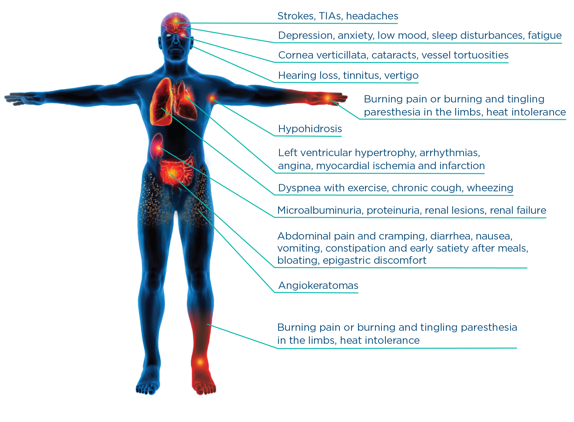
TIA=transient ischemic attack.
In a US study of 2848 patients in the Fabry Registry:
- The life expectancy in males was 58.2 years (vs. 74.7 years in the general population), while in females it was 75.4 years (vs. 80.0 years in the general population).*

If you suspect Fabry disease in one of your patients, take action.
Learn about testing options for Fabry disease
* Data from 2848 patients in the Fabry Registry were summarized using descriptive statistics. Life expectancy at birth was compared with that of the United States general population.
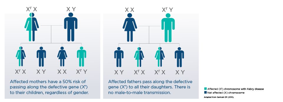
How is Fabry disease inherited?
Explaining Fabry Disease Inheritance to Patients and Families: X-Linked, Not Recessive1te
What are the clinical red flags?
Are you looking for Fabry disease?
You play an important role in identifying patients with this progressive, often life-altering genetic disease.
Clinical manifestations of Fabry disease include stroke, cardiomyopathy, and renal insufficiency6
From an online database study of 67 publications that spanned 1995–2017 and screened for people living with Fabry disease (n=51,363), the prevalence of Fabry disease in patients who present with these manifestations was:7

~0.13%
in patients with stroke
~0.18%
in patients with end-stage renal disease
What is the hallmark of Fabry disease?8
GL-3 accumulation and Alpha-Galactosidase (α-GAL A) Deficiency
- Fabry disease is caused by mutations in the galactosidase alpha (GLA) gene that cause deficiency of the enzyme α-galactosidase A (α-GAL A)8
- This results in continuous, progressive cellular accumulation of lysosomal globotriaosylceramide (GL-3) and its deacetylated form (Lyso-GL3) throughout the body8,9
- GL-3 buildup starts in utero and accumulation of GL-3 eventually leads to life-threatening complications typically in the third to fifth decade of life10
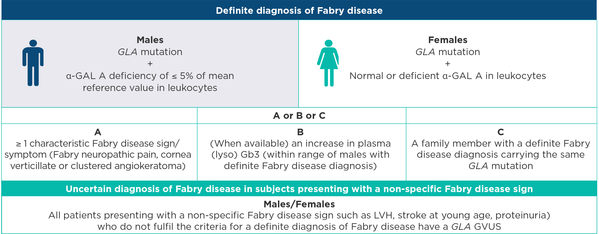
Adapted from Smid BE, et al.
α-GAL A=lysosomal α-galactosidase A enzyme; GLA mutation=defined as any abnormality found in GLA gene; LVH=left ventricular hypertrophy defined as MWT N 12 mm Gb3=globotriaosylsphingosine; GVUS=genetic variant of unknown significance, is defined as a GLA mutation that has unknown clinical significance because it does not fulfill the criteria of a definite diagnosis of Fabry disease.
In patients with an uncertain diagnosis of Fabry disease, presenting with signs such as LVH, renal failure, or proteinuria, the gold standard for a diagnosis of Fabry disease is demonstration of the characteristic in the affected organ.
Please see guidelines for complete recommendations.

Explore clinical red flags in your specialty
Recommendations for cardiac disease implications of Fabry disease12
- 2D and Doppler echocardiography is recommended in all patients at first clinical visit, with the development of new symptoms and every 12 to 24 months
- In symptomatic patients with LVH, Doppler echocardiography during exercise in the standing, sitting or semi-supine position is recommended to detect provocable LV outflow obstruction and exercise-induced mitral regurgitation
Consider cardiac MRI over echocardiography when available
Please see guidelines for complete recommendations.
LV=left ventricle; LVH=left ventricular hypertrophy.

Case study cardiomyopathies visualized by imaging techniques
Cardiomyopathies of Fabry disease include left ventricular hypertrophy (LVH) and/or hypertrophic cardiomyopathy (HCM)
Cardiac imaging may include:14
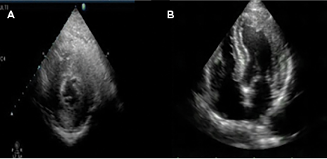
Concentric ventricular hypertrophy on echocardiography
Echocardiogram showing ventricular hypertrophy via the left chamber (A) and 4-chamber apical (B)
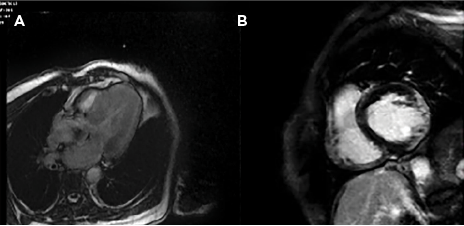
Hypertrophic cardiomyopathy on cardiac magnetic resonance
Cardiac magnetic resonance (T1) showing hypertrophic cardiomyopathy with hyperenhancement relief at the left ventricular wall.
Actual Fabry patient. May not be representative of all patients.
Adapted from Serra W and Marziliano N.
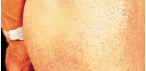
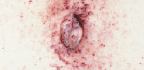
Images reproduced with permission of Robert Desnick, MD.
Angiokeratomas appear as clusters of small reddish purple, raised, non-blanchable skin lesions. They are usually found between the umbilicus and thigh in Fabry disease, with onset typically in childhood.1,2
Other cutaneous manifestations1
- Telangiectasias
- Hypohidrosis/anhidrosis
- Lymphedema
In a 2,446 patient registry, 138 patients had a stroke. It was shown that stroke was often the first major clinical event in patients with Fabry disease15*
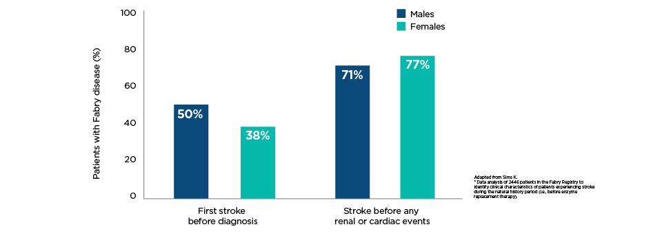
Median age of Fabry patients at first stroke15

39 Years

46 years
Risk of stroke in Fabry patients vs. general population15 (patients 35-45 years)
4.89 strokes per 1,000 person years of follow-up in males
1.83 strokes per 1,000 person years of follow-up in females
Other neurological manifestations1
- Pain
- Auditory abnormalities
- White matter lesions
Adapted from Sims K.
* Data analysis of 2,446 patients in the Fabry Registry to identify clinical characteristics of patients experiencing stroke during the natural history period (i.e., before enzyme replacement therapy).
Renal histopathology may include:
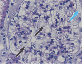
Podocyte injury
Vacuolization of mesangial cells and podocytes. The black arrows point to mesangial cells and the blue arrow points to the podocyte cell.
Adapted from Rozenfeld PA, et al.17
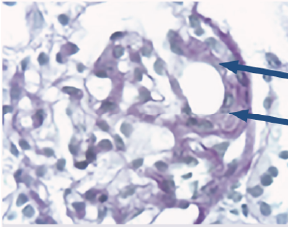
Glomerular sclerosis
Glomerulus with early signs of focal segmental glomerulosclerosis. The arrows point to the glomerulus.
Adapted from Tøndel C, et al.18
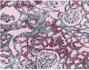
Tubulo-interstitial fibrosis
Mallory trichromic reaction showing mild interstitial fibrosis. Erythrocytes and tissue are stained red, whereas the fibrosis is stained blue.
Adapted from Rozenfeld PA, et al.17
Actual Fabry patient. May not be representative of all patients.
- Cornea verticillata
- Posterior capsular cataracts with whitish spoke-like deposits of granular material (Fabry cataract)
- Aneurysmal dilation of thin-walled venules on the bulbar conjunctiva
- Mild-to-marked tortuosity and angulation of the retinal vessels
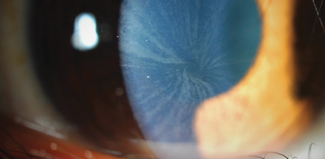
Typical cornea verticillata and corneal haze
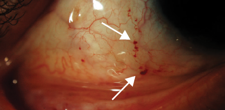
Tortuosity of the retinal vessels
Actual Fabry patient. May not be representative of all patients.
Images used with permission of Langis Michaud, OD, MSc, FAAO (Dipl).
How is Fabry disease diagnosed?
Consensus recommendations define a definite diagnosis of Fabry disease as follows:12
- GLA mutation with ≤5% GLA activity (leucocytes, mean of reference value, males only)
- ≥1 characteristic Fabry disease symptom or sign (neuropathic pain, cornea verticillata, angiokeratoma) or increased plasma (lyso) GL-3 (classical male range) or family members with definite Fabry disease
- Gold standard is defined as characteristic storage in an endomyocardial biopsy on electron microscopy
Subjects with LVH failing these criteria have GVUS and an uncertain diagnosis.
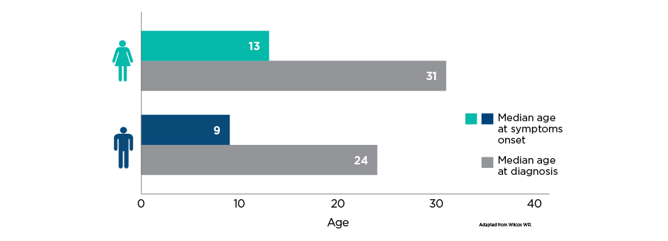
From an analysis of the US Fabry Registry patients (n=2,236) and data collection period that finalized in 2007, the median age of onset was 13 years for females and 9 years for males.
- From symptoms onset to a diagnosis of Fabry disease, there was a delay of at least 15 years in both males and females
Fabry Disease Genetic Testing Options and Workflow

Dried Blood Spot (DBS) Testing for Fabry Disease
In case of Fabry disease suspicion, you can test your patient using a dried blood spot (DBS) sample. In males, the level of α-galactosidase A (α-GAL A) enzyme activity is determined and, if low, should revert to single-gene sequencing. In females, single-gene sequencing should be performed first.
HCM panel
In the event of uncharacterized left ventricular hypertrophy (LVH), you can have your patient undergo genetic testing for hypertrophic cardiomyopathy (HCM) at your cardiovascular genetics reference centre or through external laboratory testing.1,21

GLA Gene Panel and Sequencing for Fabry Disease
DNA analysis for GLA gene mutations is necessary to confirm a diagnosis of Fabry disease in females.1,21

Family and Cascade Testing for Fabry Disease
X-linked genetic condition with a high transmission rate – testing may identify affected relatives early.1,21,22
Test the patient, test the family
For every index patient diagnosed, an average of 5 additional affected family members may be identified22
Canadian Regional Expert Centres for Fabry Disease
You can also refer your patient to the regional expert centre for the diagnosis, treatment and follow-up of Fabry disease.
Click on your region for the contact information in your province.
ATLANTIC REGIONAL EXPERT CENTRE FOR FABRY DISEASE
Halifax
Queen Elizabeth II Health Sciences Centre
Dr. Michael West – Nephrologist
Contact:
Phone: 1-877-998-9797
Fax: 902-473-8099
Email: michael.west@nshealth.ca
Centre for Clinical Research
5790 University Avenue, Rm. B06
Halifax, NS B3H 1V7
BRITISH COLUMBIA REGIONAL EXPERT CENTRES FOR FABRY DISEASE
Vancouver
Adult Metabolic Diseases Clinic
Dr. Anna Lehman
Contact:
Phone: 604-875-5965
Fax: 604-875-5967
Email: anna.lehman@vch.ca
2775 Laurel Street, 4th Floor Vancouver, BC V5Z 1 M9
Gordon and Leslie Diamond Health Care Centre
ALBERTA REGIONAL EXPERT CENTRES FOR FABRY DISEASE
Calgary
Metabolics and Genetics in Calgary (M.A.G.I.C.) Clinic
Dr. Aneal Khan
Contact:
Phone: 587-885-3158
Fax: 587-441-8380
Email: clinic@magiccalgary.ca
#102, 6715 8 Street NE, T2E 7H7
Calgary, AB
Calgary
Metabolic Genetics Clinic Alberta Children's Hospital
Dr. Kim McBride
Contact:
Phone: 403-955-2711
Fax: 403-592-4218
Email: Kim.McBride@albertahealthservices.ca
28 Oki Drive NW, T3B 6A8
Calgary, AB
Edmonton
Clinical and Metabolic Genetics Clinic Department of Medical Genetics University of Alberta
Dr. Shailly Jain
Contact:
Phone: 780-407-7333
Fax: 780-407-6845
Email: shailly.jain@albertahealthservices.ca
8-53 Medical Sciences Building
8613 114 Street, T6G 2H7
Edmonton, AB
ONTARIO REGIONAL EXPERT CENTRES FOR FABRY DISEASE
Ottawa
CHEO (Children’s Hospital of Eastern Ontario)
Dr. Danielle Bourque – Biochemical Geneticist
Contact:
Stacey Smallshaw – Nurse Case Manager
Phone: 613-737-7600 #6522
Email: ssmallshaw@cheo.on.ca
Hamilton
McMaster Children’s Hospital, Genetics Clinic
Dr. Shaymaa Shurrab – Pediatric Geneticist
Contact:
Phone: 905-521-2100 #75085
905-521-5085 (Newborn Screening Centre)
Fax: 905-521-2602
Toronto
University Health Network (UHN), Fred A. Litwin Family Centre for Genetic Medicine
Dr. Chantal Morel – Medical Geneticist
Contact:
Phone: 416-586-4800 #4220
Referral Fax: 416-619-5523
Crystal Quist, RN BScN MPH Clinical Nurse Specialist – Inherited Metabolic Diseases Program
Phone: 416-586-4800 #3083
Email: crystal.quist@uhn.ca
QUEBEC REGIONAL EXPERT CENTRES FOR FABRY DISEASE
Montreal
Hôpital Sacré-Cœur
Dr. Daniel Bichet – Nephrologist
Contact:
Nurse team
Carole Fortier – Clinical Nurse, Nephrology
Phone: 514-338-2222#3110
Email: carole.fortier.cnmtl@ssss.gouv.qc.ca
Claudia Ménard
Phone: 514-338-2222#3720
Email: claudia.menard.cnmtl@ssss.gouv.qc.ca
Kim Beauchesne
Phone: 514-338-2222#2748
Email: kim.beauchesne.cnmtl@ssss.gouv.qc.ca
Quebec City
Hôpital Hôtel-Dieu
Dr. Paul Isenring – Nephrologist
Contact:
Phone: 418-525-4444 #15464 or #15886
Email: paul.isenring@crchudequebec.ulaval.ca
Sherbrooke
Hôpital Fleurimont
Dr. Bruno Maranda – Geneticist
Contact:
Caroline Barr – Clinical Nurse, Genetics
Phone: 819-346-1110 #13933
Email: caroline.barr.ciussse-chus@ssss.gouv.qc.ca
- Germain DP. Orphanet J Rare Dis 2010;5:30. doi: 10.1186/1750-1172-5–30.
- Desnick RJ, et al. Ann Intern Med 2003;138:338–46
- Mehta A, Beck M, Sunder-Plassmann G, editors. Fabry Disease: Perspectives from 5 Years of FOS. Oxford: Oxford PharmaGenesis; 2006. Available from: https://www.ncbi.nlm.nih.gov/books/NBK11586/.
- Laney DA, et al. Genet Med 2015;17(5):323–33.
- Waldek S, et al. Genet Med 2009;11(11):790–6.
- Kolodny EH and Pastores GM. J Am Soc Nephrol 2002;13(suppl 2):S150–3.
- Doheny D, et al. J Med Genet 2018;55:261–8.
- Bokhari SRA, et al. Fabry Disease. In: StatPearls. Treasure Island, FL: StatPearls Publishing; 2022. Available from: https://www.ncbi.nlm.nih.gov/books/NBK435996/.
- Svarstad E & Marti HP. The Changing landscape of Fabry Disease. Clin J Am Soc Nephrol 2020;15(4):569–76.
- Ellaway C. Transl Pediatr 2016;5(1):37–42.
- Smid BE, et al. Int J Cardiol 2014;177:400-8.
- Linhart A, et al. Eur J Heart Fail 2020;22:1076–96.
- Linhart A and Elliott PM. Heart 2007;93:528–35.
- Serra W and Marziliano N. J Cardiovasc Ultrasound 2019;17:1.
- Sims K, et al. Stroke 2009;40:788–94.
- Terryn W, et al. Nephrol Dial Transplant 2013;28(3):505–17.
- Rozenfeld PA, et al. Mol Genet Metab 2020;129(2):132–41.
- Tøndel C, et al. Am J Kidney Dis 2008;51(5):767–76.
- Desnick RJ, Ioannou YA and Eng CM. α-Galactosidase A Deficiency: Fabry Disease. In: Valle D, et al. eds. New York, NY: McGraw-Hill; 2014. Available from: http://ommbid.mhmedical.com/content.aspx?bookid=971&Sectionid=62644837.
- Wilcox WR, et al. Mol Genet Metab 2008;93(2):112–28.
- Mehta A, et al. Q J Med 2010;103(9):641–59.
- Laney D and Fernhoff PM. J Genet Counsel 2008;17:79–83.
© 2026 sanofi-aventis Canada Inc. All rights reserved.
MAT-CA-2600656E
View-SoloSTAR®-video-(EN)
View-DoubleSTAR®-video-(EN)

View-SoloSTAR®-video-(FR)

View-DoubleSTAR®-video-(FR)
