Pathophysiology of Bullous Pemphigoid
In bullous pemphigoid, autoimmunity to BP180 and BP230 is thought to trigger a type 2 inflammatory response. Read this article to find out more

The role of type 2 inflammation in bullous pemphigoid: Exploring beyond autoimmunity
Autoimmunity to BP180 and BP230 is thought to trigger a type 2 inflammatory pathway in bullous pemphigoid1–9
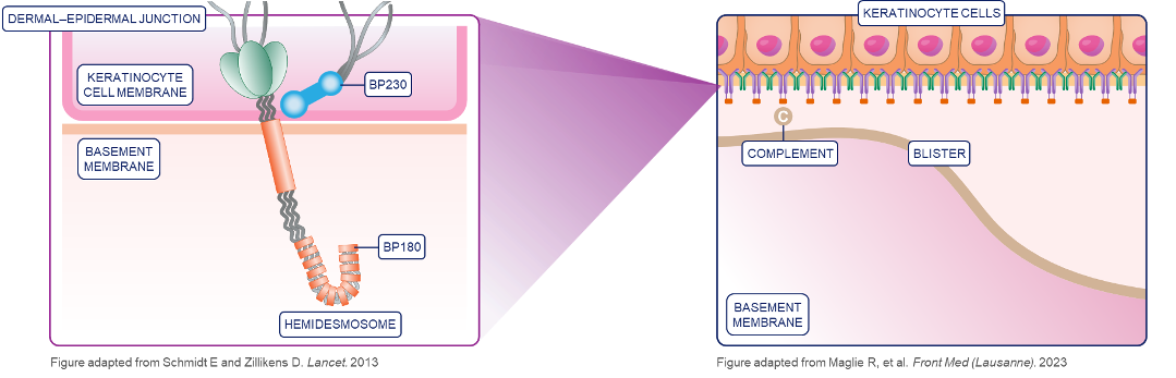
BP180 and BP230 are proteins responsible for the anchoring of keratinocytes to the basement membrane in complexes known as hemidesmosomes.1–3
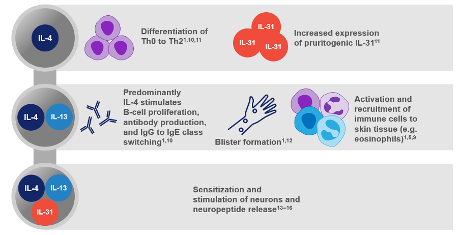
In bullous pemphigoid, naïve T cells are activated and differentiate into autoreactive T helper type 2 (Th2) cells, which secrete type 2 cytokines including interleukin (IL)-4, IL-13, and IL-31. IL-4 particularly plays a more prominent role in stimulating B-cell proliferation and immunoglobulin (Ig) G to IgE class switching.1–7
IgG and IgE autoantibodies targeting BP180 and BP230 are produced and bind to the basement membrane zone and initiate a complement cascade. In parallel, keratinocytes begin to release proinflammatory cytokines into the environment in a complement-independent manner.1,3,4,8,9
The role of type 2 inflammatory mediators in bullous pemphigoid
Type 2 inflammatory cytokines, IL-4, IL-13, and IL-31, play multiple roles in the pathophysiology of bullous pemphigoid, including blister formation.1,5,10–18
These cytokines also contribute to itch via the sensitization and stimulation of neurons and neuropeptide release.13–16
Eosinophils, basophils, and mast cells play critical roles in blister formation17,18
Eosinophil infiltration is prominent in BP, with eosinophil-secreted proteases potentially contributing to dermal-epidermal separation17
Eosinophils, basophils, and mast cells release pro-inflammatory mediators, including IL-4 and IL-13, which also contribute to blister formation17,18
Itch in bullous pemphigoid may have multiple mediators and potentially indicates a type 2 inflammatory response13–15,18,19
Intense itch is one of the primary symptoms of bullous pemphigoid, with 85% of patients suffering from itch daily20,21
Contributors to itch in bullous pemphigoid include eosinophils, basophils, and mast cells which release potent itch inducers, including IL-4, IL-13, IL-31 and histamine13–15,18,19
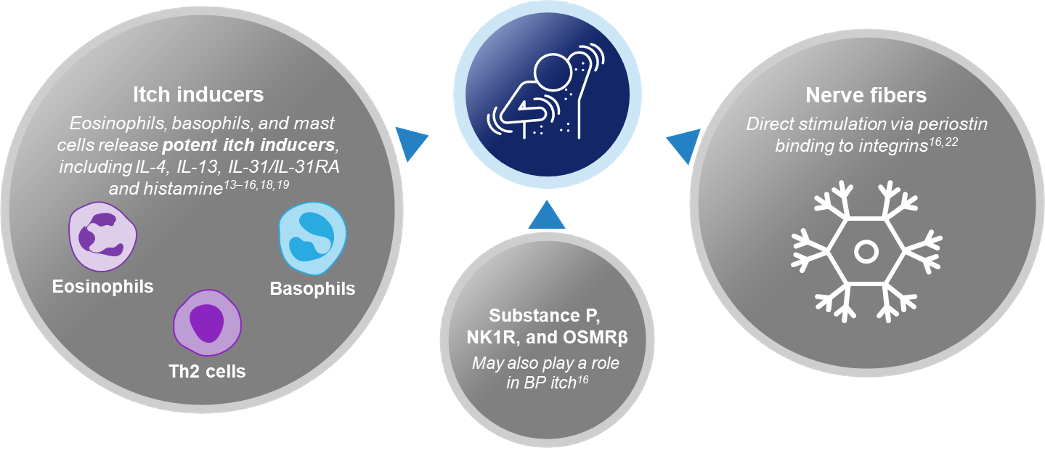
Evidence suggests that itch severity in bullous pemphigoid is directly linked to periostin deposition (which directly stimulates nerve fibers), number of eosinophils, basophils, IL-13(+) cells and IL-31(+) cells16,22
|
What is bullous pemphigoid?
|
The challenges of managing bullous pemphigoid
|
Morbidity and mortality in bullous pemphigoid
|
Current treatment approaches and unmet needs in bullous pemphigoid
|
- Zhang L, et al. Front Immunol. 2023;14:1115083.
- Muramatsu K, et al. J Allergy Clin Immunol. 2018;142(6):1818–30.e6.
- Zhang J, et al. J Invest Dermatol. 2018;138(9):1917–24.
- Ujiie H. J Dermatol. 2023;50(2):140–9.
- Thoma-Uszynski S, et al. J Immunol. 2006;176:2015–2.
- Ujiie H, et al. J Immunol. 2010;184:2166–74.
- Schinner J, et al. Front Immunol. 2023;14:1203776.
- Nelson KC, et al. J Clin Invest. 2006;116:2892–900.
- Cole C, et al Front Immunol. 2022;13:912876.
- Fang H, et al. Autoimmun Rev. 2020;19(11):102661.
- Stott B, et al. J Allergy Clin Immunol. 2013;132:446–454.
- Huang R, et al. Ann Med. 2023;55:2280991.
- Gibbs BF, et al. Front Immunol. 2019;10:1383.
- Ingrasci G, et al. Exp Dermatol. 2021;30(9):1208–17.
- Rüdrich U, et al. Acta Dermato-Venereologica. 2018;98:766–71.
- Hashimoto T, et al. J Am Acad Dermatol. 2020;83:53–62.
- Amber KT, et al. Front Med. 2018;5:201.
- Limberg MM, et al. Biomolecules. 2023;13:1019.
- Hashimoto T, et al. Exp Dermatol. 2019;28:1373–79.
- Schmidt E, et al. J Am Acad Dermatol. 2002;47:133–6.
- Briand C, et al. Acta Derm Venereol. 2020;100:adv00320.
- Hashimoto T, et al. J Invest Dermatol. 2021;141:2338–43.

Considerations for bullous pemphigoid management
The management of bullous pemphigoid is challenging and new treatment approaches are needed. Read this article to find out more.

Patient morbidity and mortality in BP
Many factors, including older age and multimorbidity, are associated with mortality in bullous pemphigoid. Read this article to find out more

What is Bullous Pemphigoid
The heterogeneous presentation of bullous pemphigoid may shed light on why patients are underdiagnosed. Read the article to learn more