cGVHD criteria
cGvHD is characterized by a combination of tissue inflammation and fibrosis
Global grading of cGvHD1
The diagnosis and assessment of cGVHD are guided by the National Institutes of Health (NIH) Consensus Criteria, which provide a standardized framework for identifying, staging, and managing the disease. This section outlines the global severity grading of cGVHD, guidance on diagnosis, and diagnostic characteristics of cGvHD.
| No of organs involved | Mild Grade | Moderate Grade | Severe Grade |
|---|---|---|---|
| 1 | Score 1 | Score 2 | Score 3 |
| 2 | Score 1 | Score 2 | Score 3 |
| 3 | Score 1 | Score 3 | |
| Lung | Score 1 | Score 2 |
Moderate grade: Lung score 1 or ≥3 organs at score 1 or at least one organ at score 2.
Severe grade: Lung score 2 or score 3 in any organ.
 SKIN
SKIN MOUTH
MOUTH EYES
EYES GI TRACT
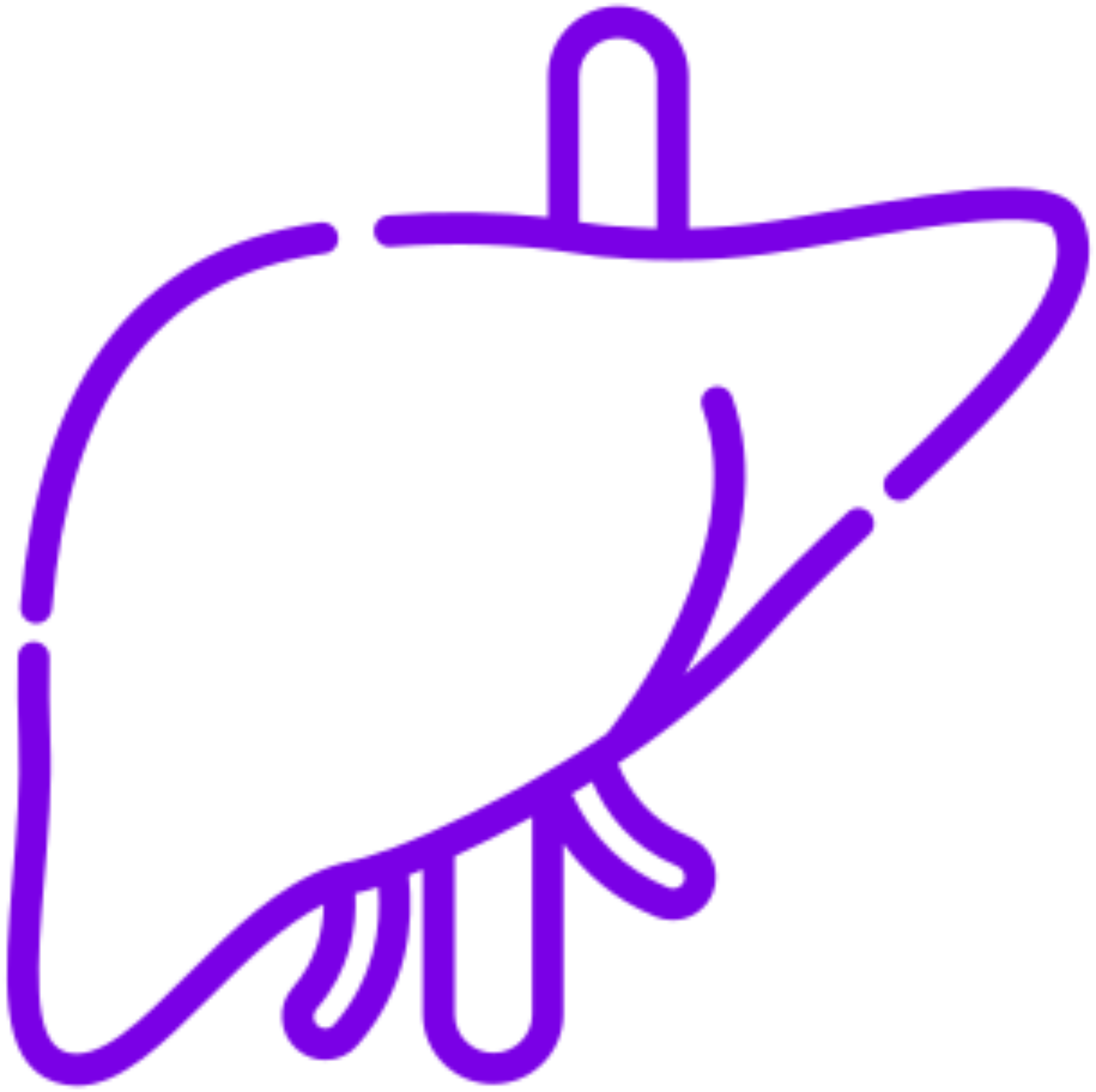 LIVER
LIVER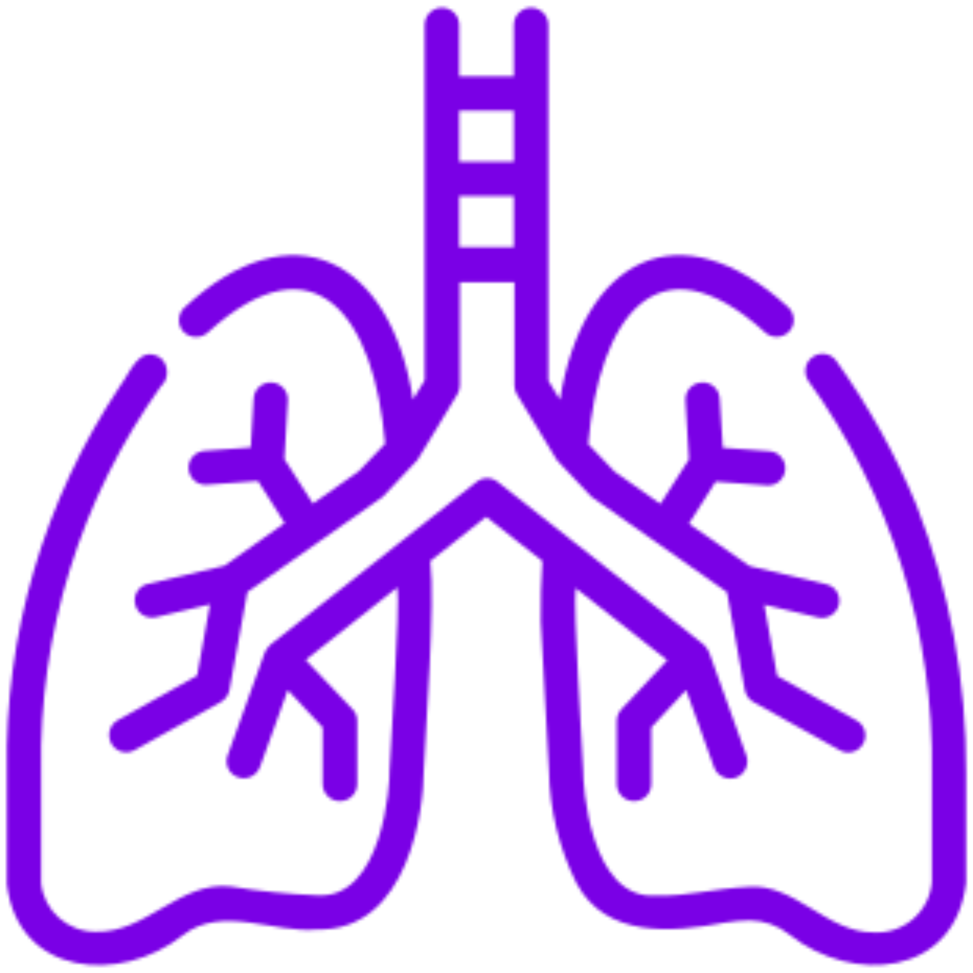 LUNGS
LUNGS JOINTS AND FASCIA
 GENITAL TRACT
- Immune cells and their target tissues produce various cytokines and chemokines, which cause inflammation and tissue damage.7
- The sclerotic skin manifestations of cGvHD (ScGvHD) result from inflammation and fibrosis of the dermis, subcutaneous tissue, or fascia, leading to significant functional disability.8
- Sclerotic fibrosis of the perioral tissue owing to chronic inflammation can lead to restricted oral range-of-motion in patients with oral cGvHD.9
- Conjunctival inflammation may be a primary manifestation of cGvHD or secondary to severe dry eye.10
- The intestine can be the target of several immunologically mediated diseases, including cGvHD and inflammatory bowel disease.11
- Pulmonary cGvHD leads to BOS which results in obstructive and/or restrictive changes.12
- Esophageal involvement in cGvHD results in mucosal inflammation, leading to submucosal fibrosis and, occasionally, formation of webs and strictures.13
- Liver cGvHD usually presents as an indolent cholestatic disease in patients with skin, mouth, and eye involvement.14
Guidance on diagnosis in the 2014 NIH cGvHD consensus criteria15
NIH categorizes clinical features as
- Diagnostic
- Distinctive
- Other features or unclassified manifestations
- Common
A diagnosis of cGvHD requires the presence of ≥1 diagnostic features OR ≥1 distinctive features plus additional testing.
(e.g., biopsy, laboratory tests, evaluation by a specialist, radiographic imaging)
Diagnostic
Clinical features that establish the presence of cGvHD and do not require additional testing or evidence of other organ involvement
Distinctive
Clinical features that are not typically found in aGvHD and do require additional testing (e.g., biopsy) to establish a diagnosis of cGvHD
Other features or unclassified
Rare, controversial or nonspecific characteristics of cGvHD that cannot be used to establish a diagnosis
Common
Clinical features shared by cGvHD and aGvHD
Diagnostic characteristics of cGvHD
To provide structure and standardization in diagnosing cGvHD, particularly in the clinical trial setting, the NIH Consensus Development Project on Criteria for Clinical Trials in cGvHD published the 2014 guidance on diagnosing and staging, commonly referred to as the 2014 NIH cGvHD Consensus Criteria.15
The clinical features of cGvHD are grouped into 4 categories: diagnostic, distinctive, other features or unclassified manifestations and common.15
Prior to the 2014 NIH cGvHD Consensus Criteria, the characteristics of cGvHD were classified as limited or extensive.16
The 2014 NIH cGvHD consensus criteria also guide assessment of the severity of cGvHD
SKIN
MOUTH
EYES
GI TRACT
LIVER
LUNGS
JOINTS AND FASCIA
GENITAL TRACT
- Chronic GvHD is classified into mild, moderate or severe, based on a global score
- The global score takes into consideration the involvement and severity of 8 organs or sites
- Each organ or site score ranges from 0 (no involvement) to 3 (severe impairment
Key points
|
aGvHD, acute graft-versus-host disease; BOS, Bronchiolitis Obliterans Syndrome; cGvHD, chronic graft-versus-host disease; GI, gastrointestinal; GvHD, graft-versus-host disease; NIH, National Institutes of Health.
- Kim D.D.H et al. Curr Oncol. 2024;31:1426-1444;
- Kitko CL et al. Biol Blood Marrow Transplant. 2012;18(1 suppl):S46-S52. doi:10.1016/j.bbmt.2011.10.021;
- Cooke KR et al. Biol Blood Marrow Transplant. 2017;23(2):211-234. doi:10.1016/j.bbmt.2016.09.023;
- MacDonald KPA et al. Blood. 2017;129(1):13-21. doi:10.1182/blood-2016-06-686618;
- Fiuza-Luces C et al. Bone Marrow Transplant. 2016;51(1):13-26. doi:10.1038/bmt.2015.195;
- Henden AS, Hill GR. J Immunol. 2015;194(10):4604-4612. doi:10.4049/jimmunol.1500117;
- Presland RB. World J Transplant. 2016;6(4):608-619. doi.org/10.5500/wjt.v6.i4.608;
- Martires KJ et al. Blood. 2011;118(15):4250-7. doi.org/10.1182/blood-2011-04-350249;
- Bassim CW et al. J Dent Res. 2015;94(4):547-54. doi.org/10.1177%2F0022034515570942;
- Ogawa Y et al. Sci Rep. 2013;3:3419. doi.org/10.1038%2Fsrep03419;
- Haring E et al. Front Immunol. 2021;12:705342. doi.org/10.3389/fimmu.2021.705342;
- Flowers MED, Martin PJ. Blood. 2015;125(4):606-615. doi:10.1182/blood-2014-08-551994;
- Schima W et al. Abdom Imaging. 1994;19(3):191-4. 10.1007/BF00203504;
- Strasser SI et al. Hepatology. 2000;32(6):1265-71;
- Jagasia MH et al. Biol Blood Marrow Transplant. 2015;21(3):389-401.e1. doi:10.1016/j.bbmt.2014.12.001.


Bevestig uw status
Bevestig uw status
Sanofi biedt u exclusieve content over onze verschillende therapeutische gebieden op één portal
speciaal voor professionals in de gezondheidszorg, inclusief wetenschappelijk nieuws, een ljst met
evenementen en tools en diensten voor uw dagelijkse praktik.